
Proper Use & Care of Microscopes Clinician's Brief
The cells for distinguishing levels of intensity, not color, are the rod cells. Each of these cell types are located on the retina at the back of the inside of the eye. The front of the eye (see Figure 2), which includes the iris, the curved cornea, and the lens, admits light and focuses it on the retina.
Anatomy and Physiology I Coursework Microscope A+P
No matter what you love, you'll find it here. Search Microscopes Parts and more. Looking for Microscopes Parts? We have almost everything on eBay.
Parts Parts And Functions Of A Microscope
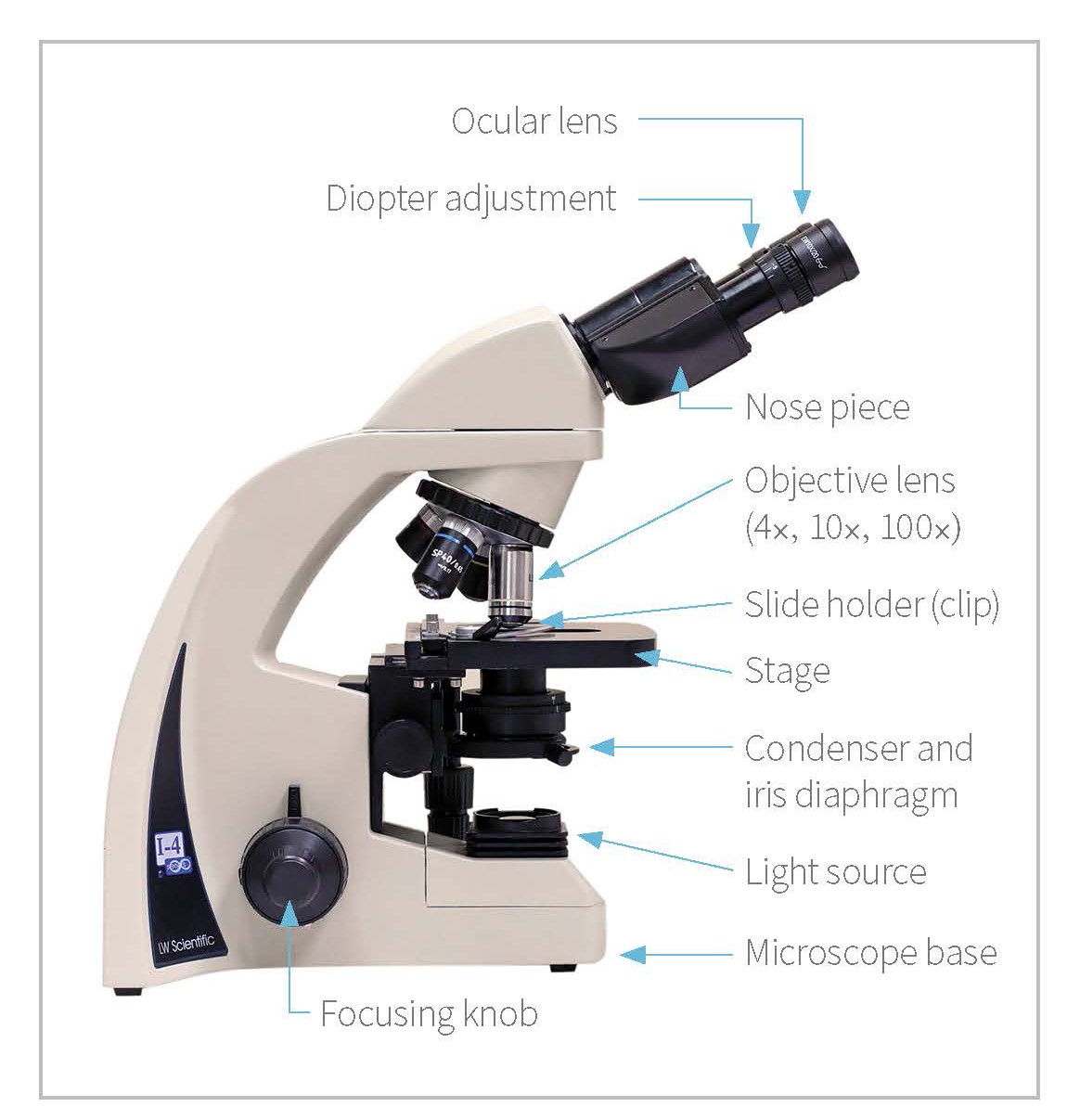
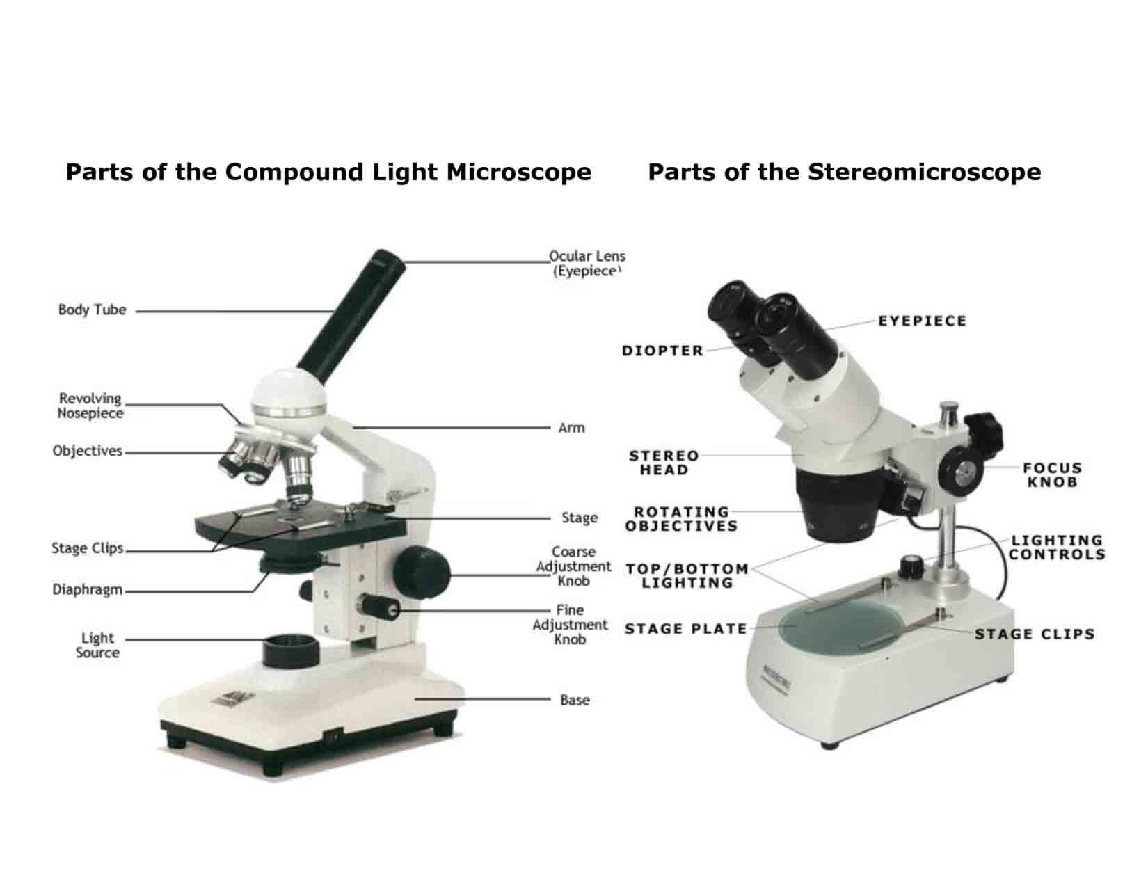
Power switch Condenser Parts of Compound Microscope (Labeled Pictures) a. Mechanical Parts of a Compound Microscope Foot or Base Pillar Arm Stage Inclination Joint Clips Diaphragm Nose piece/Revolving Nosepiece/Turret Body Tube Adjustment Knobs b. Optical Parts of a Compound Microscope Eyepiece lens or Ocular Mirror Objective Lenses

Microscope Diagram Labeled, Unlabeled and Blank Parts of a Microscope
Having been constructed in the 16th Century, microscopes have revolutionized science with their ability to magnify small objects such as microbial cells, producing images with definitive structures that are identifiable and characterizable. Derived from Greek words "mikrós" meaning "small" and "skópéō" meaning "look at". Table of Contents
Microscopes 7th Grade Science
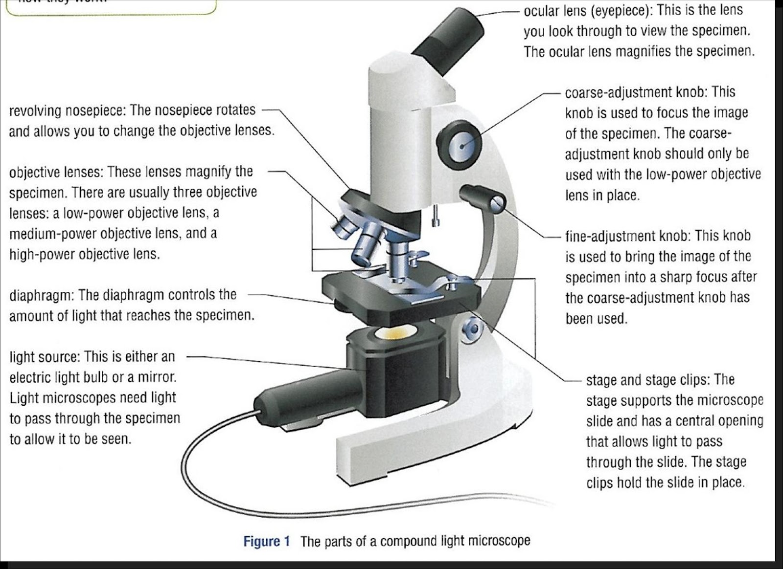
Image 2: The body tube part of a microscope is where the ray of light is bent to allow the object being viewed to enlarge by the scope. Picture Source: slideplayer.com 3. Turret/Nose piece. It is the revolving part of the microscope. It allows the use of different types of objective lenses by simply rotating the top part of the turret.
microscope parts and functions 571 plays Quizizz
Category: Science & Tech Key People: Ernst Abbe John Henry Dallmeyer Joseph Jackson Lister Jean de Hautefeuille Antonie van Leeuwenhoek (Show more) Related Topics: electron microscope acoustic microscope X-ray microscope electron microscopy (Show more)
DIY Home Shatnez Lab Part 2 How to Use the Monocular Compound Student
Microscopy Introduction to microscopes and how they work. Covers brightfield microscopy, fluorescence microscopy, and electron microscopy. Introduction If you meet some cell biologists and get them talking about what they enjoy most in their work, you may find it comes down to one thing: secretly, they're all microscope freaks.

Microscope Diagram Labeled, Unlabeled and Blank Parts of a Microscope
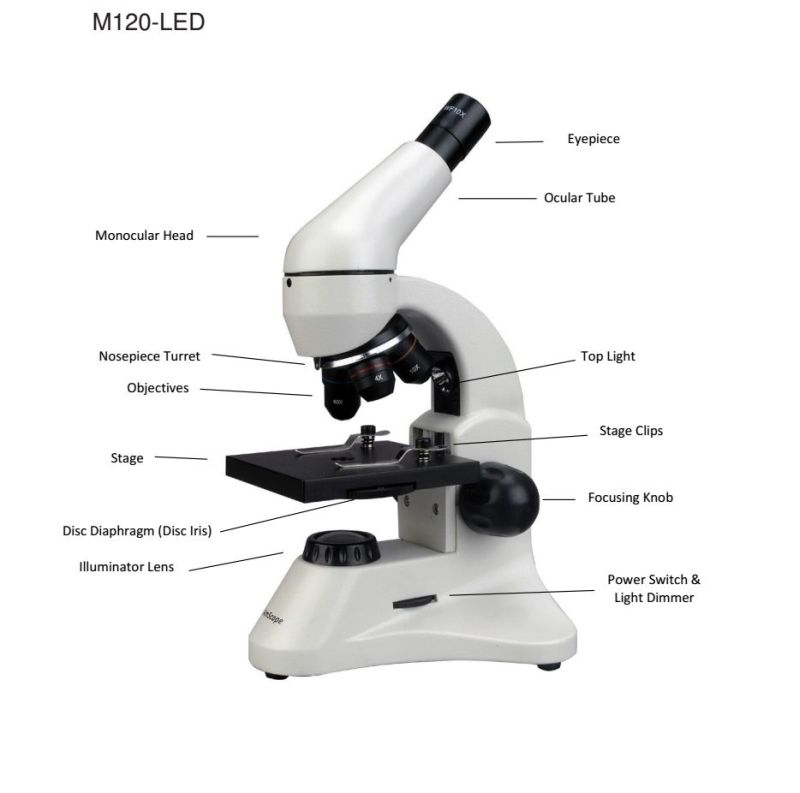
What are the Parts of a Microscope? Eyepiece Lens: the lens at the top that you look through, usually 10x or 15x power. Tube: Connects the eyepiece to the objective lenses. Arm: Supports the tube and connects it to the base. Base: The bottom of the microscope, used for support.
The Microscope Its Parts and Their Functions
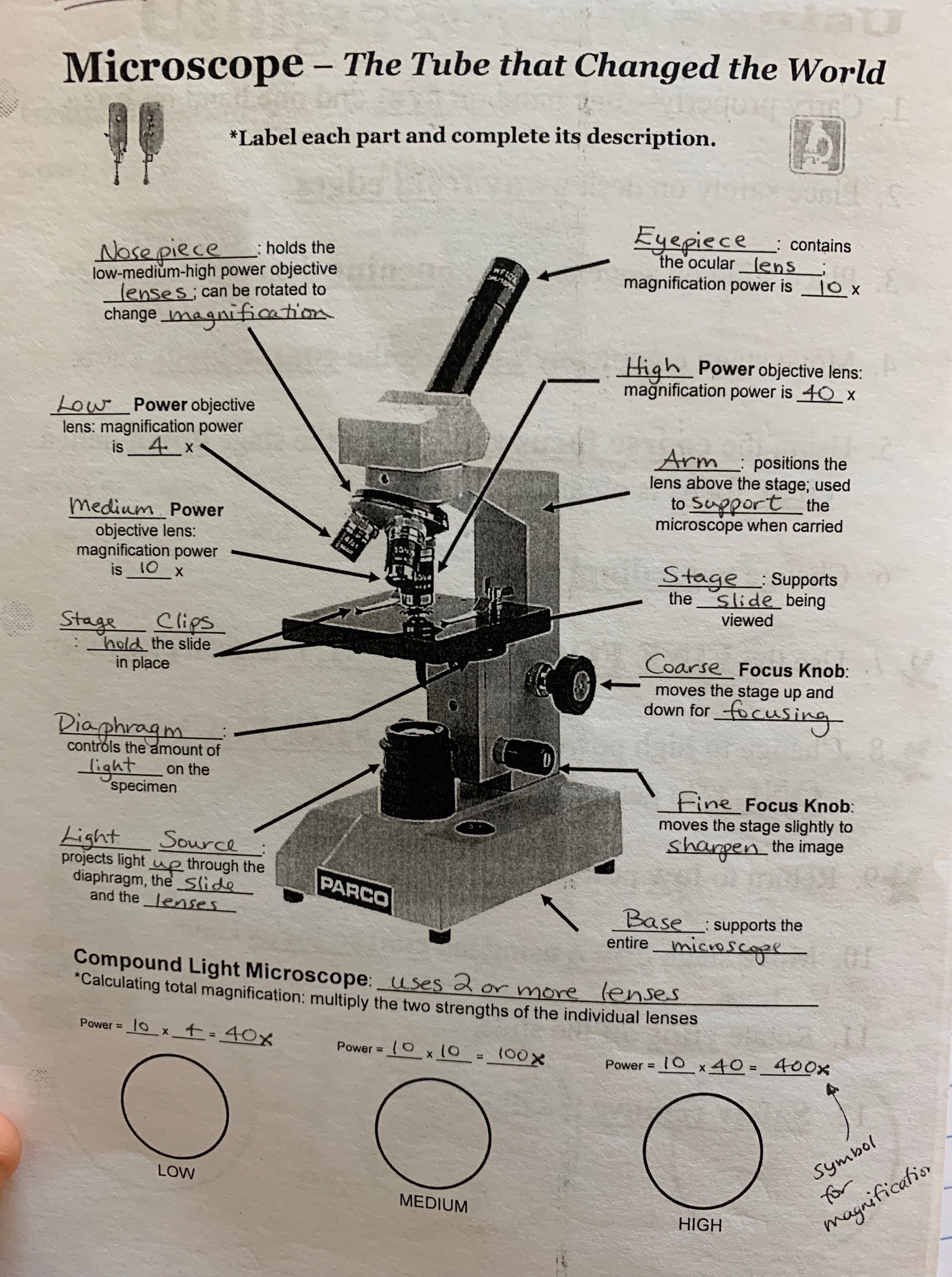
Parts of a microscope. A compound microscope uses two or more lenses to produce a magnified image of an object, known as a specimen, placed on a slide (a piece of glass) at the base. The microscope rests securely on a stand on a table. Daylight from the room (or from a bright lamp) shines in at the bottom.
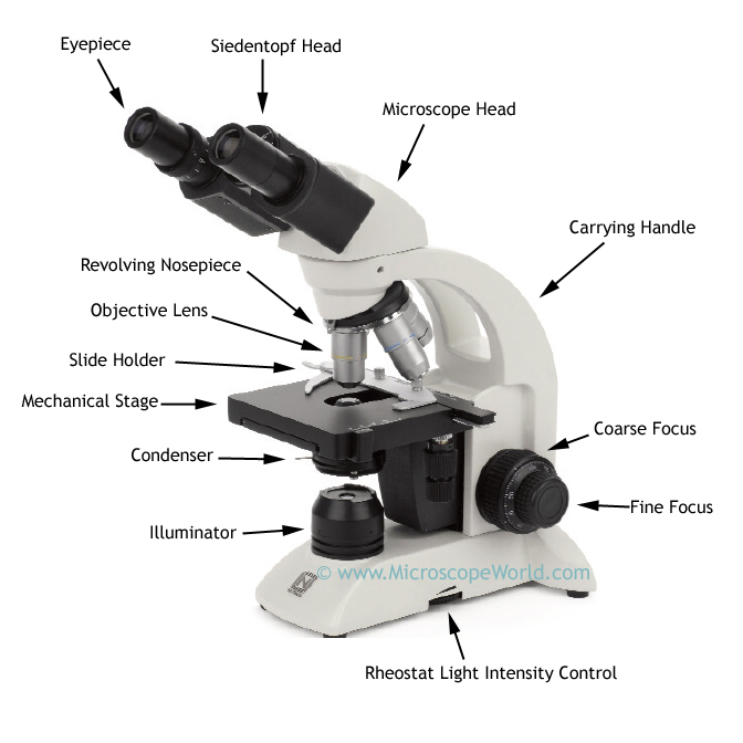
Microscope World Blog Biological Microscope Parts
Parts of a Microscope with Their Functions Written by Sushmita Baniya in Lab Equipment Last Updated December 30, 2023 A microscope is a piece of laboratory optical equipment used to magnify small things that are too small for the details to be seen by the naked eye.
Light Microscope Main Parts Of Light Microscope Biology —
SEM (Scanning Electron Microscope) - It scans through the surface of the specimen by focusing the electron beam. As a result of technical advancements, one can also find more efficient microscopes like scanning probe microscopes and scanning acoustic microscopes. Labelled Microscope Parts Compound Microscope with Parts Name

Microscope Diagram to Print 101 Diagrams
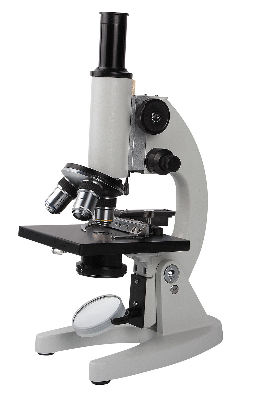
Figure 2: Brightfield light microscope used in a Microbiology lab (Lumen) The Optical System. The optical system of a compound microscope consists of two lens systems: one found in the objective(s) lens(es) (Fig. 2, part 3); the other in the ocular (eyepiece) (Fig. 2 part 1).

Mrs. Conner's Science Place Microscope Parts
Body tube (Head): The body tube connects the eyepiece to the objective lenses. Arm: The arm connects the body tube to the base of the microscope. Coarse adjustment: Brings the specimen into general focus. Fine adjustment: Fine tunes the focus and increases the detail of the specimen. Nosepiece: A rotating turret that houses the objective lenses.

The Top 5 Microscopes for Kids (And How to Use Them!) Spring Into STEM
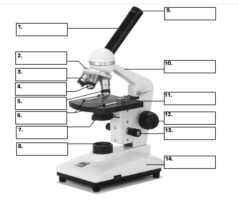
All microscopes share features in common. In this interactive, you can label the different parts of a microscope. Use this with the Microscope parts activity to help students identify and label the main parts of a microscope and then describe their functions.. Drag and drop the text labels onto the microscope diagram. If you want to redo an answer, click on the box and the answer will go back.

36 Label Parts Of The Microscope Labels 2021
Eyepiece Tube The eyepiece tube is another essential microscope part. This is where the various lenses work together and subsequently do their magic. Not only do you look down it to get a view at the stage and see a magnified view of the specimen you're examining, but it is what makes a microscope work.

🎉 Main components of a light microscope. Parts of a microscope with
There are several components to the modern light microscope that come together to enhance its function: The base of the microscope provides stability to the device and allows the user's hands to be free to manipulate other aspects of the microscope or document relevant observations.